
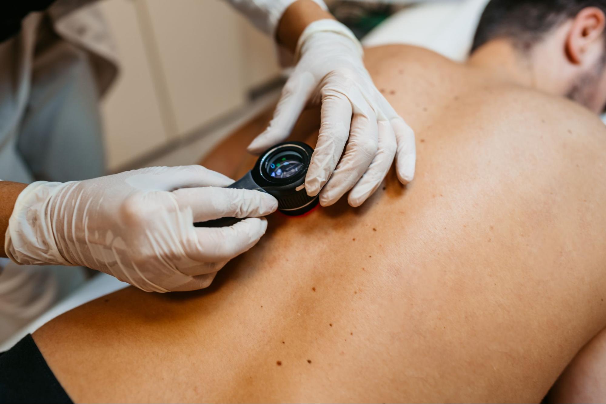

Mohs surgery is a focused procedure used in both dermatologic surgery and pathology; it treats certain types of skin cancer through a staged process. A physician removes a thin layer of tissue, examines it under a microscope, and repeats the process only where abnormal cells remain. Unlike some other surgical approaches, tissue removal and examination occur during the same visit. Here’s more information about Mohs surgery:
Defining the Procedure
Mohs surgery treats selected skin cancers by removing tissue in carefully mapped layers. After each layer is removed, the physician examines the edges and underside of that tissue under a microscope. This step-by-step review helps identify the exact location of any remaining abnormal cells. Unlike a standard excision, Mohs surgery does not rely on broad removal followed by delayed lab review; it uses immediate microscopic analysis at the treatment site. This structure makes the process distinct and highly methodical. The procedure joins surgery, mapping, and slide interpretation into one coordinated workflow.
Explaining the Main Steps
The procedure may begin with the preparation of the treatment area and the use of local anesthesia. Once the area is numb, the physician removes the visible tumor and a very thin layer of surrounding tissue. The removed sample is marked and color-coded; it is also mapped to match its exact position on the skin.
The tissue then moves to an on-site lab area for processing. A technician prepares microscope slides from the sample, and the physician reviews them to check for remaining abnormal cells at the margins. If any cells appear, the map shows their location. The physician then removes another thin layer only from that marked area. This cycle continues until the examined margins show no remaining abnormal cells.
Identifying Common Uses
Physicians use Mohs surgery for specific skin cancers, especially basal cell carcinoma and squamous cell carcinoma. They may also use it in selected cases involving other skin tumors; this depends on the diagnosis and clinical setting. Use varies by:
- Lesion type
- Size
- Location
- Prior treatment history
Describing Tissue Preservation
A key feature of Mohs surgery is tissue preservation. Because the physician removes additional tissue only where abnormal cells remain, the procedure is designed to avoid unnecessary removal of surrounding skin. This makes the technique especially relevant in areas where preserving structure is fundamental to function or appearance, such as the:
- Nose
- Eyelids
- Lips
- Ears
- Hands
- Scalp
This targeted approach also shapes how the surgical site is managed after the final stage. Once the margins are clear on microscopic review, the physician addresses the wound. Closure methods vary. The wound is stitched closed in some situations; others require a skin flap, a graft, or natural healing, depending on the site and the treatment plan.
Outlining Time Requirements
Mohs surgery usually takes place in an outpatient setting. The tissue removal itself is sometimes brief, but slide preparation and microscopic review require added time between stages. Full visits may last several hours. The total length depends on how many stages are needed.
Learn More About Mohs Surgery
The Mohs surgical technique removes certain skin cancers while examining tissue during the same appointment. It involves mapped tissue removal, on-site microscopic review, and repeated stages only when abnormal cells remain. The procedure timing and repair method vary by case, but the overall structure remains consistent. To learn more about this surgery, contact a qualified specialist today.