
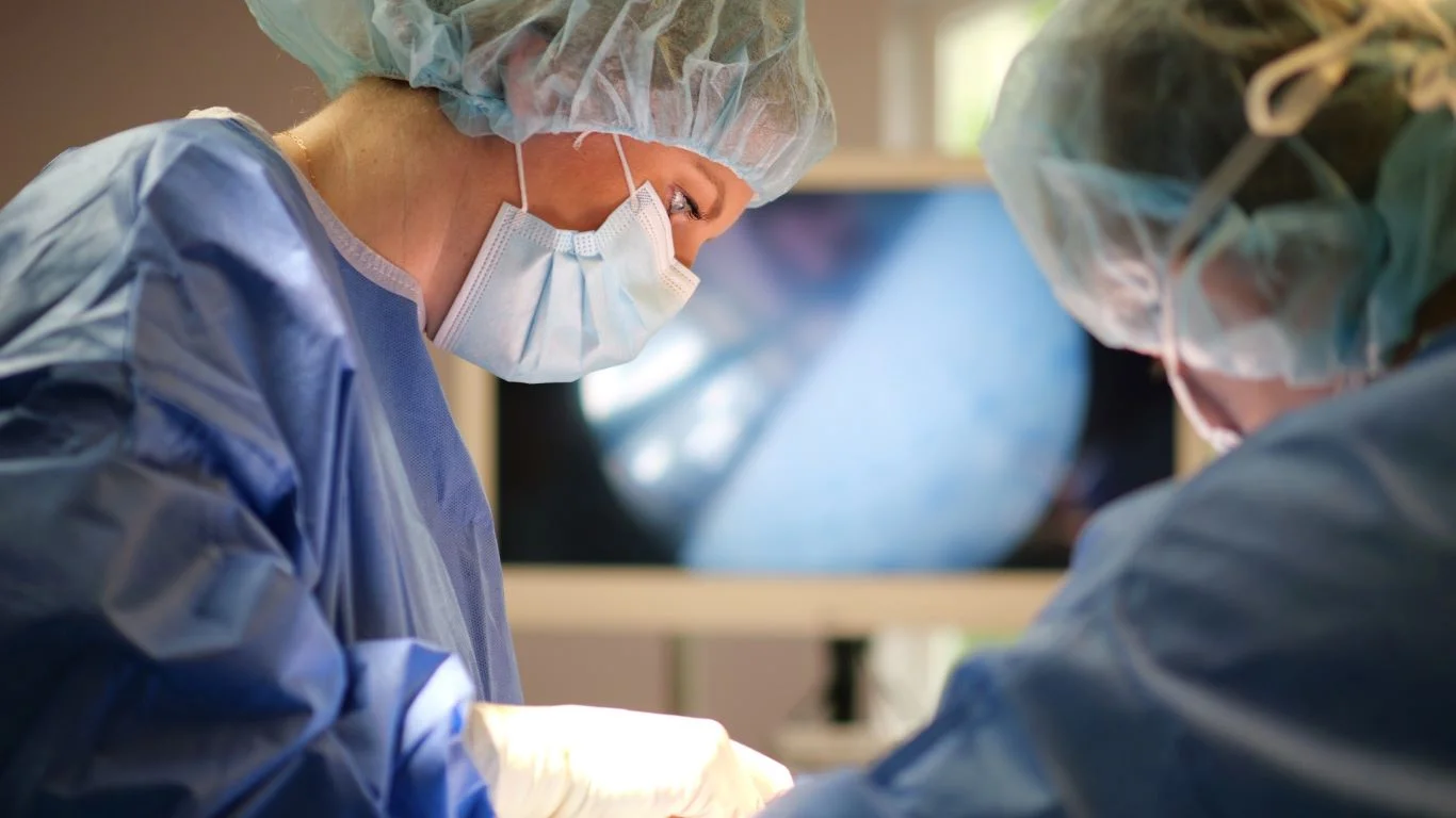

-When you have stomach issues, endoscopy is a medical procedure that lets doctors examine the digestive tract directly. A thin, flexible tube fitted with a camera is guided through the mouth or rectum. This tool gives physicians a clear, real-time view of internal tissue, and it helps pinpoint problems that imaging scans miss. Here is more information on the role of endoscopy in diagnosing digestive disorders:
Examining the Esophagus and Stomach
The esophagus connects your throat to your stomach, and both organs are common sites of digestive disease. Doctors insert the endoscope through your mouth to reach these areas. Since the camera transmits live images, the physician sees the tissue condition immediately. Endoscopy of the upper digestive tract covers several key areas:
- The lining of the esophagus
- The valve between the esophagus and stomach
- The stomach walls and lining
- The upper part of the small intestine
Tissue samples, called biopsies, are collected during the same procedure. The physician uses small instruments passed through the endoscope, and lab results from these samples confirm or rule out specific conditions. Abnormalities that appear suspicious under the camera prompt immediate sampling.
Using an Endoscope
An endoscope is a long, flexible tube roughly the width of a finger. It carries a light source and a high-definition camera at its tip. The physician steers the tube with precision, and the images appear on a monitor in real time. Different attachments allow for tissue sampling, fluid removal, or minor surgical tasks.
Two types of endoscopy serve distinct diagnostic purposes:
- Upper endoscopy (EGD): examines the esophagus, stomach, and upper small intestine
- Colonoscopy: examines the large intestine and rectum
When the physician needs detailed images, the camera zooms into suspicious tissue. Narrow-band imaging highlights blood vessel patterns that signal early cellular changes. This technology adds a layer of detail to standard visual examination.
Detecting Ulcers
Peptic ulcers are open sores that form on the stomach lining or upper small intestine. Endoscopy locates these sores with accuracy, and the physician notes their size, depth, and appearance. Since ulcers vary widely in severity, direct visualization guides treatment decisions more precisely than symptom reports alone.
The physician also checks for signs of bleeding during the same examination. Active bleeding from an ulcer appears as a visible blood source on the monitor. Endoscopy allows for immediate intervention if bleeding is detected, which reduces the need for separate surgical procedures.
Biopsies taken from ulcer sites test for bacteria linked to peptic ulcers. The lab analyzes tissue samples, and results direct the physician toward targeted treatment. Identifying the bacteria early narrows the diagnostic path significantly.
Diagnosing Acid Reflux
Acid reflux occurs when stomach acid moves upward into the esophagus repeatedly. Over time, this exposure damages the esophageal lining. Endoscopy reveals the degree of that damage by showing tissue inflammation, erosions, or a condition called Barrett’s esophagus, where normal cells are replaced by abnormal ones.
Doctors measure the extent of tissue changes using standardized classification systems. Grading the severity informs the physician’s next steps, and documented findings create a baseline for follow-up examinations. Patients with certain issues require regular endoscopic monitoring because cellular changes progress at variable rates.
Schedule Endoscopy Sessions Near You
Endoscopy provides direct, detailed information about the digestive tract that other diagnostic tools cannot replicate. It identifies ulcers, tissue damage, and cellular abnormalities with specificity and speed. Contact a gastroenterology clinic today to schedule your endoscopy consultation. Bring a record of your symptoms, their duration, and any medications you take to your appointment.